Ultrastructural pathology for airway defence mechanism

University of Cambridge – MRC Toxicology Unit, UK
Nobuhiro Morone
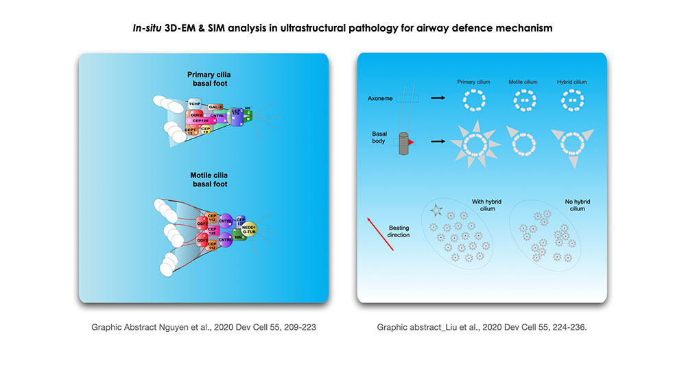
In-situ three-dimensional analysis of intracellular organelles and protein complexes is essential to understand the role of individual components and their cellular function. Through 3D-SIM (Structural Illumination Microscopy), Vito Mennella’s group characterized the organization of the ciliary basal foot, an appendage of basal bodies whose main role is to provide a point of anchoring to the microtubule cytoskeleton (Nguyen QPH et al., Dev Cell 2020; Liu et al., Dev Cell 2020). They visualized that the basal foot is organized into three main regions linked by elongated coiled-coil proteins, revealing a conserved modular architecture in primary and motile cilia, and identified CEP112 as a basal foot protein and other candidate components of the assembly. In this collaborative project, we will address an ultrastructural molecular architecture under EM resolutions by In-situ cryo-tomography to understand deeply the role of basal foot body.